
제품소개
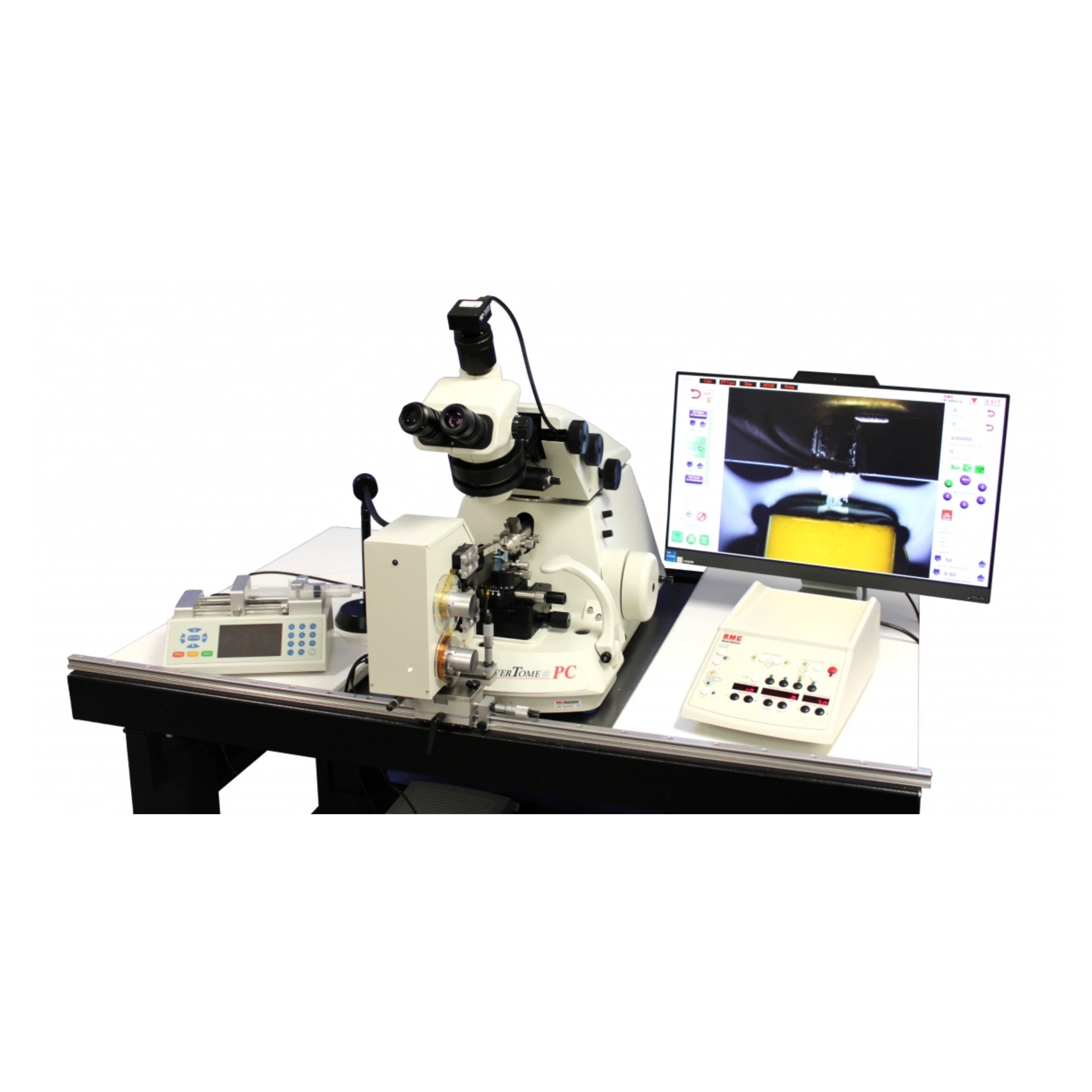
ATUMtome
- ATUM continuous tape feed mechanism with PC touchscreen control
- PowerTome PTPCZ with HD Video Package
- Air-activated anti-vibration table with Compressor and Ergonomic lab chair
- Diatome Ultra Maxi Diamond Knife, 4mm, 35°
- Start-up supply of Kapton tape
- 4” diameter silicon wafers
- Environmental chamber
- Anti-static device
- Water level control system
- Wafer workstation
상세정보
ATUMtome: Automated Tape Collecting Ultramicrotome
The expansion and advancement of microscopical applications has led to exciting new developments, not least in the field of 3D Array Tomography and Volume Imaging.
At Harvard University, Professor Jeff Lichtman, PhD, MD, and his team designed an instrument (ATUM) capable of collecting very large sets of serial sections (30,000+) in an unattended and computer-controlled operation, to help reconstruct the brain’s neural pathways — a precursor to one day mapping the entire human brain. The ATUM is connected to and controlled by RMC Boeckeler’s PowerTome ultramicrotome, to form the ATUMtome (Automatic Tape Collecting Ultramicrotome).
While developed initially for the neurosciences, the versatile nature of serial collection combined with the precise and robust capabilities of the PowerTome, has seen the use of the ATUMtome expand into other research areas.
- Collects hundreds to thousands of sections on a continuous tape
- Non-destructive to sample, with sections available for years into the future for processing, post-staining, immunogold labeling, correlative imaging — and at any work pace desired
- Section thickness can range from 0nm to 15μm
- Determines sample viability sooner in the process so you don’t waste valuable time cutting and imaging a problem sample. Uses multiple resolutions for locating regions of interest, then zooming in for higher resolution imaging
- Allows correlative microscopy for localization and then ultra-structural imaging
- Prepares samples that allow short pixel dwell time for fast detection of images during electron microscopy
- Uses standard sample preparation techniques and resins
- Charging effect on sections is manageable
- Priced at a fraction of the cost of alternative 3-D imaging techniques